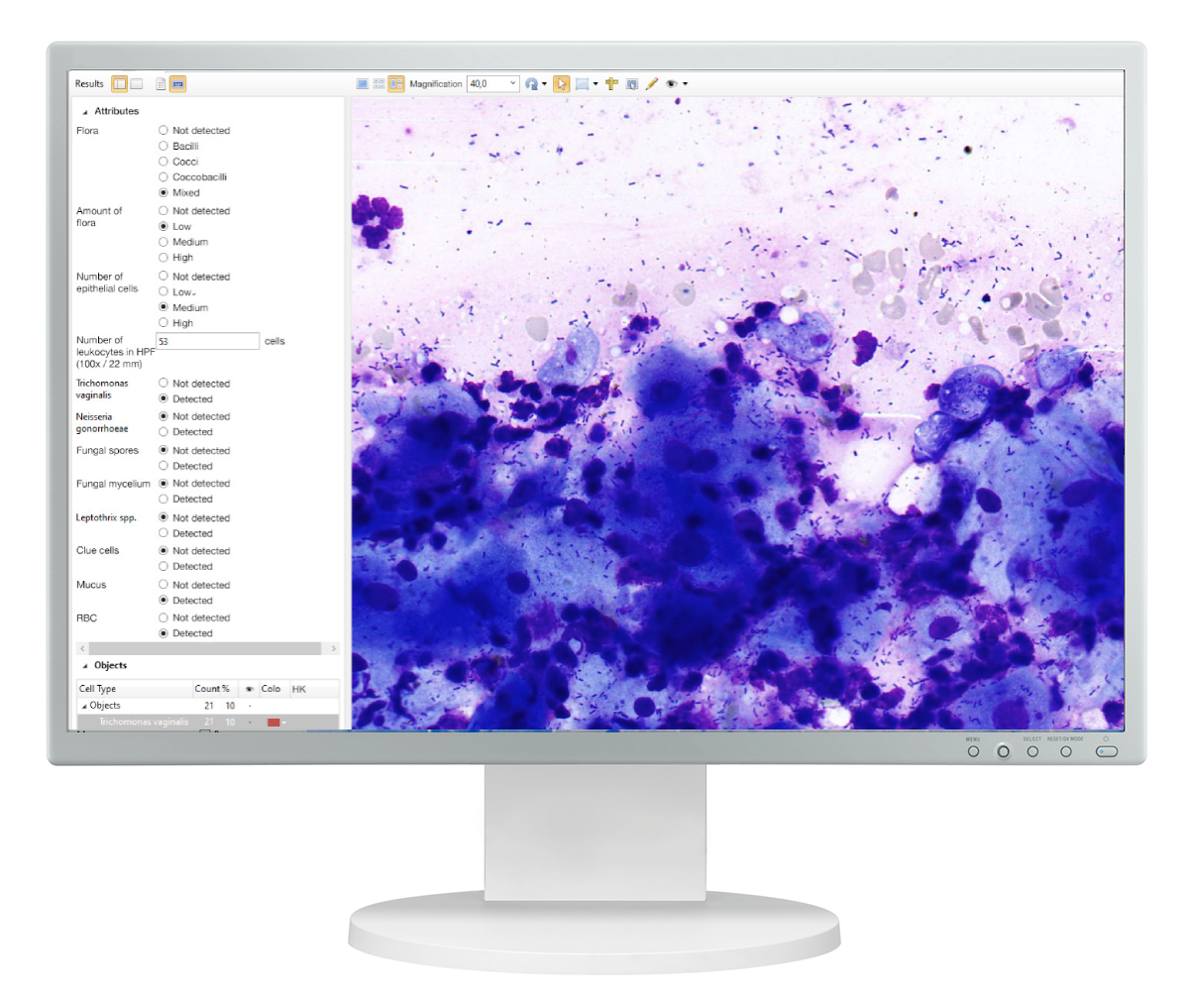
Vision Cyto STD (Sexually Transmitted Diseases)
Microbiome
CST
Vaginal smear
Microscopic examination (Cervical, Urethral, Vaginal)
Microscopy for STI pathogens
Microscopy for N.gonorrhoeae
Classification parameters and objects
— 1/3 of CUV localization
— Type and amount of flora
— Epithelium
— Leukocytes
— Trichomonas vaginalis
— Neisseria gonorrhoeae
— Fungal spores
— Fungal mycelium
— Leptothrix spp
— Clue cells
Sample images from the cell gallery

Trichomonas vaginalis

Neisseria gonorrhoeae

Fungal spores

Fungal mycelium

Leptothrix spp.
Download
West Medica | STD
*Product specifications and terms of use may vary by region and are subject to change without prior notice